
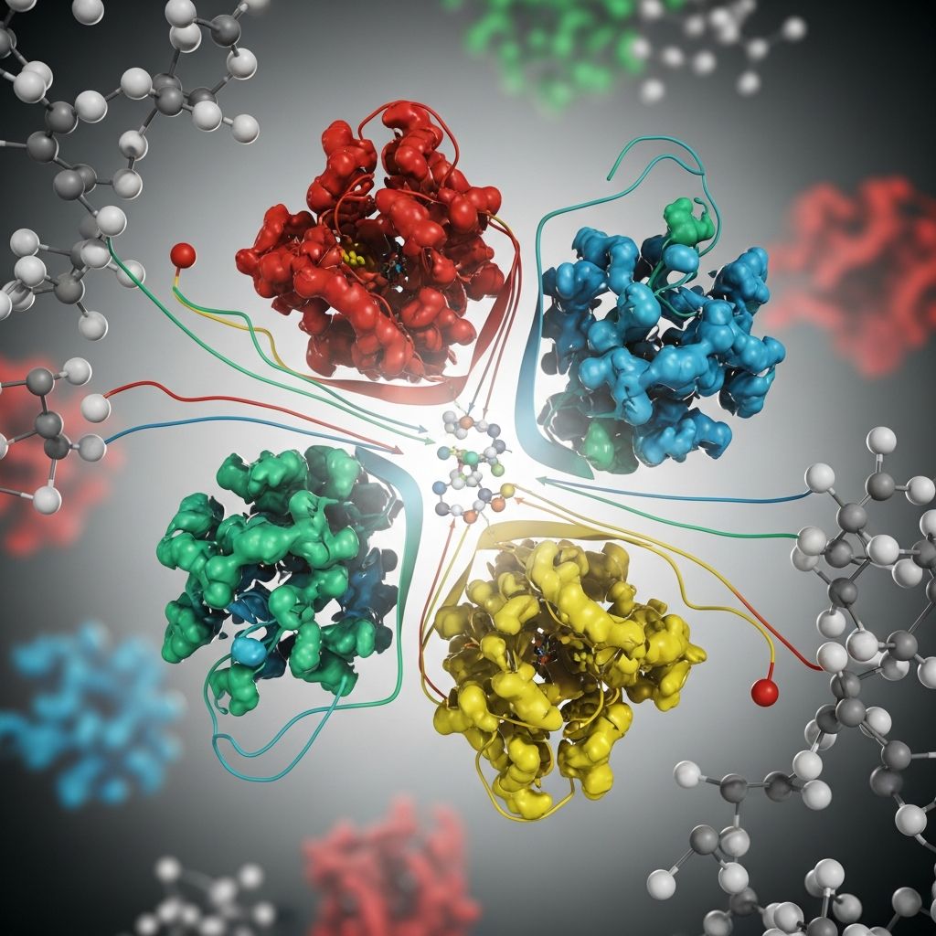
Enzymes serve as the workhorses of cellular metabolism, accelerating chemical reactions essential for life. These proteins, and occasionally RNA molecules, possess highly specialized structures that enable precise substrate binding and catalysis without being consumed in the process.
The Molecular Architecture of Enzymes
At their core, enzymes are polypeptides—chains of amino acids folded into complex three-dimensional shapes. The primary structure, defined by the linear sequence of amino acids, dictates all higher levels of organization. This sequence folds into secondary structures like alpha-helices and beta-sheets, stabilized by hydrogen bonds, and further assembles into tertiary structures through hydrophobic interactions, disulfide bridges, and ionic bonds.
Many enzymes function as single polypeptide units, while others form quaternary structures with multiple subunits. For instance, fatty acid synthase comprises thousands of amino acids across multiple chains, showcasing the diversity in enzyme size and complexity. The globular nature of most enzymes positions their functional regions on the surface, optimizing interactions with substrates.
Key Structural Features: Active Site and Beyond
The **active site** is the enzyme’s catalytic heart—a cleft or pocket formed by specific amino acid residues. This region binds substrates through complementary shapes and chemical properties, often involving non-contiguous amino acids brought together by the protein’s fold.
Adjacent to the active site lie **binding sites** that orient substrates precisely for reaction. The rest of the enzyme scaffold maintains this site’s geometry and dynamics. Some enzymes incorporate **allosteric sites**, where regulatory molecules bind to modulate activity via conformational changes.
- Catalytic residues: Directly participate in bond breaking/forming.
- Binding residues: Stabilize substrates via hydrogen bonds, electrostatics, or van der Waals forces.
- Structural domains: Provide rigidity and flexibility for function.
Dynamics: Enzymes in Motion
Far from rigid locks, enzymes exhibit dynamic fluctuations. Internal motions—ranging from side-chain wobbles to domain shifts—create a conformational ensemble that facilitates catalysis. These vibrations lower activation energy barriers by positioning catalytic groups optimally.
X-ray crystallography reveals flexible loops near active sites, with high temperature factors indicating mobility. In cyclophilin A, such dynamics drive peptidyl-prolyl isomerization, coupling structure, motion, and function. Solvent interactions further influence these movements, with hydration shells providing energy for barrier crossing.
Substrate Binding Models
| Model | Description | Key Features |
|---|---|---|
| Lock and Key | Substrate fits active site perfectly like a key in a lock. | Rigid enzyme; complementary shapes and chemistries. |
| Induced Fit | Enzyme and substrate mold together upon binding. | Dynamic adjustment optimizes transition state stabilization. |
The induced fit model predominates, explaining enzyme specificity and versatility. Binding induces conformational changes, aligning catalytic residues and straining substrate bonds toward the transition state.
Catalytic Mechanisms and Activation Energy
Enzymes accelerate reactions by reducing **activation energy** (E_a)—the barrier to bond rearrangement. Substrates collide randomly, but enzymes bind them in reactive orientations, stabilizing the high-energy transition state.
Mechanisms include:
- Acid-base catalysis: Proton donation/acceptance by residues like histidine.
- Covalent catalysis: Transient nucleophilic attack forming enzyme-substrate intermediates.
- Metal ion catalysis: Stabilization via ions like Zn^{2+}.
The Michaelis-Menten equation models this: E + S ⇌ ES → E + P. At low [S], rate ∝ [S]; at saturation, V_max is reached.
Cofactors and Coenzymes: Essential Partners
Apoenzymes (protein only) often require non-protein helpers:
- Cofactors: Inorganic ions (e.g., Mg^{2+}, Fe^{2+}) stabilizing charge or geometry. DNA polymerase needs Zn^{2+}.
- Coenzymes: Organic molecules (e.g., NAD^+, derived from vitamins) shuttling groups like electrons.
- Holoenzyme: Complete, active complex.
Ribozymes, RNA catalysts, represent non-protein exceptions, though rare.
Regulation of Enzyme Activity
Cells fine-tune enzymes via:
- Allosteric regulation: Effectors bind distant sites, altering active site affinity.
- Covalent modification: Phosphorylation/dephosphorylation toggles activity.
- Compartmentalization: Localization in organelles controls access.
- Inhibitors: Competitive (block active site), non-competitive (alter structure).
Feedback inhibition prevents overproduction, as in metabolic pathways.
Environmental Influences on Enzyme Performance
**Temperature:** Optimal range maximizes activity; excess causes denaturation, unfolding active sites.
**pH:** Affects ionization of catalytic residues; deviations disrupt function (e.g., pepsin at low pH, trypsin at high).
**Ionic strength:** Modulates electrostatic interactions.
Enzymes in Biotechnology and Medicine
Engineered enzymes drive industry: proteases in detergents, amylases in biofuels. In medicine, enzyme replacement therapies treat deficiencies (e.g., Gaucher’s disease), while inhibitors target pathogens (statins for cholesterol).
Directed evolution and rational design optimize enzymes for non-natural substrates, expanding synthetic biology.
Future Directions in Enzyme Research
Advances in cryo-EM and AI predict structures from sequences, enabling novel catalyst design. Understanding dynamics promises efficiency gains in biocatalysis and drug discovery.
Frequently Asked Questions (FAQs)
What is the role of the active site in enzymes?
The active site binds substrates and catalyzes reactions by lowering activation energy through precise orientation and chemical facilitation.
How do enzymes achieve specificity?
Complementary shape, charge, and hydrophobicity ensure only correct substrates bind effectively, enhanced by induced fit.
Can enzymes be denatured?
Yes, heat, pH extremes, or chemicals disrupt folding, abolishing activity; renaturation is sometimes possible.
What are ribozymes?
Ribozymes are catalytic RNAs, challenging the protein-only view of enzymes, involved in splicing and peptide bond formation.
How do inhibitors affect enzymes?
Competitive inhibitors mimic substrates, blocking access; non-competitive alter structure, reducing efficacy.
References
- Enzyme — Wikipedia. 2023-10-01. https://en.wikipedia.org/wiki/Enzyme
- Enzymes – Structure, Classification, and Function — BYJU’S. 2023-01-15. https://byjus.com/biology/enzymes/
- Enzymes: An integrated view of structure, dynamics and function — PMC (NCBI). 2006-02-01. https://pmc.ncbi.nlm.nih.gov/articles/PMC1379655/
- 8.2: Enzyme Structure and Function — Biology LibreTexts. 2023-08-20. https://bio.libretexts.org/Courses/City_College_of_San_Francisco/Introduction_to_Microbiology_(Liu_et_al.)/08:_Microbial_Metabolism_I/8.02:_Enzyme_Structure_and_Function
- Enzymes — Monash University. 2024-03-10. https://www.monash.edu/student-academic-success/biology/regulation-of-biochemical-pathways/enzymes
- Biochemistry, Proteins Enzymes — StatPearls (NCBI). 2023-07-04. https://www.ncbi.nlm.nih.gov/books/NBK554481/

Medha Deb is an editor with a master's degree in Applied Linguistics from the University of Hyderabad. She believes that her qualification has helped her develop a deep understanding of language and its application in various contexts. Medha specializes in the areas of beauty, health, sports, and wellness and is committed to ensuring that the content on the website is…