
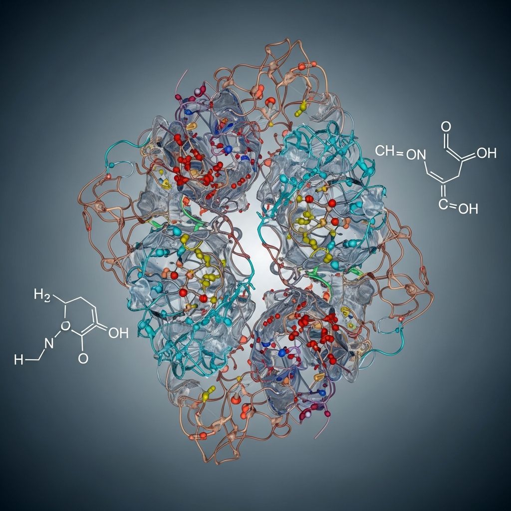
Proteins are the workhorses of cellular processes, performing tasks from catalyzing reactions to providing structural support. Their functionality hinges on precise three-dimensional shapes, which are maintained by a variety of chemical bonds. These interactions range from strong covalent linkages to weaker non-covalent forces, each contributing uniquely to protein folding and stability.
Foundation of Proteins: Amino Acids and Peptide Bonds
At the core of every protein lies a chain of amino acids linked by **peptide bonds**, the primary covalent connections that form the polypeptide backbone. Each amino acid consists of a central alpha carbon attached to a hydrogen atom, a carboxyl group (-COOH), an amino group (-NH2), and a variable side chain (R group) that defines its chemical properties.
The formation of a peptide bond occurs through a dehydration reaction, where the carboxyl group of one amino acid reacts with the amino group of another, releasing a water molecule. This creates a covalent amide linkage between the carbonyl carbon (C=O) of the first amino acid and the nitrogen (N-H) of the second. The resulting bond exhibits partial double-bond character due to resonance, restricting rotation and giving it a planar, rigid structure with a bond length of approximately 1.32 Å—intermediate between single (1.49 Å) and double (1.27 Å) bonds.
This rigidity is crucial for maintaining the extended conformation of the protein backbone, setting the stage for higher-order structures. Without peptide bonds, proteins could not form the linear polymers essential for folding into functional shapes. In a typical protein, hundreds of these bonds connect 50 to thousands of amino acids, creating a robust primary structure.
Disulfide Bonds: The Covalent Stabilizers
Among covalent interactions beyond peptide bonds, **disulfide bonds** stand out as powerful cross-links formed exclusively between cysteine residues. Cysteine’s side chain contains a thiol group (-SH), which can oxidize to form a covalent S-S bridge when two such groups are brought into proximity during protein folding.
These bonds are particularly vital in extracellular proteins, where they confer resistance to unfolding under harsh conditions. For instance, in insulin, disulfide bonds link different parts of the chain, locking the hormone into its active conformation. The formation is catalyzed in the endoplasmic reticulum by enzymes that pair -SH groups from adjacent cysteines in the folded state.
Disulfide bridges enhance tertiary and quaternary structures by reducing entropy in the unfolded state, making the native fold thermodynamically favorable. Their strength—comparable to peptide bonds—makes them irreversible under physiological conditions, providing long-term stability.
Non-Covalent Forces: Hydrogen Bonds in Protein Architecture
**Hydrogen bonds** are weaker non-covalent interactions (typically 1.5-2.5 Å) that play a dominant role in defining secondary structures like alpha helices and beta sheets. They form when a hydrogen atom bonded to an electronegative atom (N or O) interacts with another electronegative atom’s lone pair.
In an alpha helix, the carbonyl oxygen (C=O) of one residue hydrogen-bonds to the amide hydrogen (N-H) four residues ahead, stabilizing the coiled structure. Linus Pauling’s pioneering work highlighted their role in protein folding. Beta sheets involve inter-strand hydrogen bonds between backbone atoms of adjacent strands, creating pleated sheets that are strong and rigid.
Beyond secondary structures, hydrogen bonds occur between side chains, backbone atoms, and even water molecules at the protein surface. They contribute about 20-40 kJ/mol per bond, collectively stabilizing the fold against thermal denaturation.
- Intra-chain: Stabilize helices and loops.
- Inter-chain: Link beta strands in sheets.
- Side chain-backbone: Fine-tune local conformations.
Ionic Bonds: Electrostatic Attractions in Protein Folding
**Ionic bonds**, or salt bridges, arise between oppositely charged side chains, such as the negatively charged carboxylate of aspartic or glutamic acid and the positively charged ammonium of lysine or arginine.
These interactions are potent in the hydrophobic protein core, where they can rival covalent bond strength due to low dielectric constants. For example, in the enzyme chymotrypsin, a salt bridge between aspartate and a terminal ammonium group anchors structural domains. Ionic bonds are pH-sensitive; protonation or deprotonation disrupts them, explaining why protein stability varies with environmental pH.
In tertiary structure, salt bridges connect distant parts of the chain, while in quaternary structures, they mediate subunit interactions. Charge-dipole interactions with water also aid solubility of polar surfaces.
Hydrophobic Interactions: Driving the Fold
The most significant non-covalent force in protein folding is the **hydrophobic effect**, where non-polar side chains (e.g., leucine, valine, phenylalanine) cluster in the protein interior to minimize contact with water.
This is not a true bond but an entropic effect: water molecules around hydrophobes form ordered cages, reducing entropy. Burying hydrophobes releases these waters, increasing overall entropy and stabilizing the folded state. Hydrophobic cores comprise up to 50% of a protein’s interior in globular proteins.
In membrane proteins, hydrophobic interactions anchor transmembrane helices in lipid bilayers. Mutations disrupting these clusters often lead to misfolding diseases like cystic fibrosis.
Van der Waals Forces: Subtle Stabilizers
**Van der Waals attractions** are weak, transient interactions between all atoms, arising from dipole fluctuations. Effective at close range (3-4 Å), they fill spaces in densely packed cores.
Though individually feeble (1-5 kJ/mol), thousands per protein provide cumulative stability. They complement hydrophobic packing, ensuring tight atomic fits essential for specificity in enzyme active sites.
Hierarchical Organization of Protein Structures
Proteins fold hierarchically: primary structure (peptide sequence) dictates secondary elements (helices, sheets) via hydrogen bonds, which pack into tertiary folds stabilized by all forces above, and quaternary assemblies for multi-subunit proteins.
| Structure Level | Key Bonds | Function |
|---|---|---|
| Primary | Peptide bonds | Linear sequence |
| Secondary | Hydrogen bonds | Local folds (helix, sheet) |
| Tertiary | All non-covalent + disulfides | Globular 3D shape |
| Quaternary | Non-covalent interfaces | Multi-subunit complexes |
Real-World Implications: From Enzymes to Diseases
Understanding these bonds reveals why proteins function: enzymes rely on precise active site geometry from hydrogen and ionic bonds; hemoglobin’s oxygen binding involves quaternary shifts. Disruptions cause amyloid diseases (misfolded aggregates) or prions, where hydrophobic exposures drive pathology.
Drug design targets these interactions—e.g., inhibitors mimicking substrates via hydrogen bonds. Recent advances in cryo-EM visualize bond networks at atomic resolution, aiding rational protein engineering.
Frequently Asked Questions (FAQs)
What is the strongest bond in proteins?
Covalent peptide and disulfide bonds are the strongest, providing the backbone and permanent cross-links.
How do hydrogen bonds contribute to protein stability?
They define secondary structures and numerous weak interactions that collectively resist unfolding.
Why are hydrophobic interactions crucial for folding?
They drive burial of non-polar residues, providing the major free energy for the native fold.
Can ionic bonds form inside proteins?
Yes, salt bridges are common in cores, enhanced by low water exposure.
What role do disulfide bonds play in extracellular proteins?
They lock structures against proteases and denaturation in oxidizing environments.
References
- Types of Chemical Bonds in Proteins — University of Mustansiriyah. 2021-04-10. https://uomustansiriyah.edu.iq/media/lectures/6/6_2021_04_10!04_16_00_PM.pdf
- Molecular Biology of the Cell: The Shape and Structure of Proteins — NCBI Bookshelf (Alberts et al.). 2002 (authoritative classic). https://www.ncbi.nlm.nih.gov/books/NBK26830/
- Bonds Stabilizing Protein Structure — Dhing College. 2020-10-04. https://dhingcollegeonline.co.in/attendence/classnotes/files/1602006237.pdf
- Bonds and Protein Structure — California Lutheran University. N.D. (Accessed 2026). https://earth.callutheran.edu/Academic_Programs/Departments/BioDev/omm/jsmolnew/bonding/chymo.html
- Proteins – Chemistry LibreTexts — LibreTexts (Sacramento City College). 2023. https://chem.libretexts.org/Courses/Sacramento_City_College/SCC:_Chem_309_-_General_Organic_and_Biochemistry_(Bennett)/Text/16:_Proteins_and_Enzymes/16.04:_Proteins

Medha Deb is an editor with a master's degree in Applied Linguistics from the University of Hyderabad. She believes that her qualification has helped her develop a deep understanding of language and its application in various contexts. Medha specializes in the areas of beauty, health, sports, and wellness and is committed to ensuring that the content on the website is…