
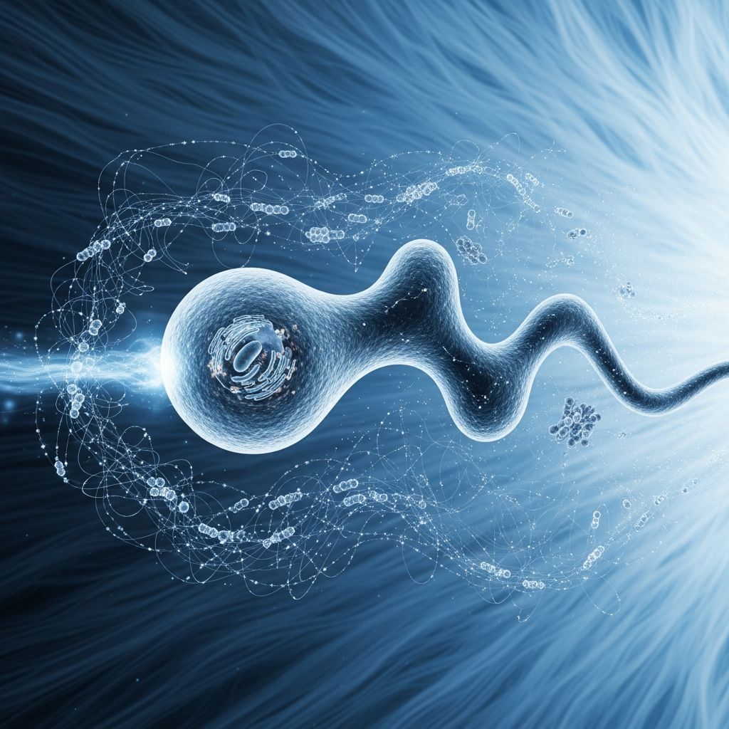
Cell motility represents a fundamental biological process enabling single cells and cell collectives to traverse their environments, underpinning essential functions from embryonic development to immune surveillance. Driven primarily by the dynamic actin cytoskeleton, this movement involves coordinated protrusion, adhesion, and contraction phases that allow cells to respond to environmental cues with precision.
Foundational Components of Cellular Locomotion
At the heart of cell motility lies the cytoskeleton, a dynamic network of protein filaments that provides structural support and generates mechanical forces. The actin cytoskeleton, composed of globular actin (G-actin) monomers assembling into filamentous actin (F-actin), serves as the primary engine for protrusion at the cell’s leading edge. Actin polymerization pushes the plasma membrane forward, creating structures like lamellipodia (broad, sheet-like extensions) and filopodia (finger-like projections).
Microtubules and intermediate filaments play supportive roles. Microtubules, radiating from the microtubule-organizing center, guide intracellular transport and contribute to polarity establishment, while intermediate filaments offer mechanical resilience during translocation. Motor proteins such as myosin II interact with actin to produce contractile forces, retracting the cell rear and propelling the body forward.
Step-by-Step Dynamics of Cell Movement
Cell migration unfolds in a cyclic process divided into three interdependent stages, each powered by cytoskeletal rearrangements and physical forces.
- Protrusion: Upon sensing directional cues, the cell initiates actin polymerization at the front. Branched actin networks, nucleated by the Arp2/3 complex, drive membrane extension. This phase transitions the cortical actin from a gel-like state to a more fluid sol, facilitating rapid advancement.
- Adhesion: Integrin-mediated focal adhesions anchor the leading edge to the extracellular matrix (ECM). These dynamic structures link the cytoskeleton to the substrate, transmitting traction forces. Optimal adhesion strength balances forward pull without excessive drag.
- Contraction and Retraction: Myosin II motors contract the acto-myosin network, generating retrograde flow and pulling the cell body forward. Simultaneously, deadhesion at the rear, often via calpain-mediated proteolysis, releases trailing edges.
These stages operate continuously, with gel-sol transitions enabling cytoskeletal fluidity. In fast-moving cells like fish keratocytes, protrusion and translocation rates synchronize seamlessly.
Environmental Signals Triggering Motility
Cells do not move randomly; they respond to gradients of chemical (chemoattractants) or mechanical stimuli detected by surface receptors. Chemotaxis guides immune cells toward infection sites, while mechanotaxis influences fibroblasts in wound healing. Receptor activation triggers signaling cascades, polarizing the cell by enriching actin regulators at the front and myosin at the rear.
Phosphoinositides like PI(3,4)P2 exemplify regulatory checkpoints. Accumulating at the rear, PI(3,4)P2 modulates Ras activity, preventing front-end hyperactivity and ensuring directional persistence. Disruption leads to erratic, inefficient migration.
| Signal Type | Examples | Cellular Response |
|---|---|---|
| Chemical | Chemokines, growth factors | Actin polymerization, polarity establishment |
| Mechanical | Substrate stiffness, shear stress | Durotaxis, enhanced adhesions |
| Electrical | Ion gradients | Galvanotaxis in neurons |
Diverse Modes of Cellular Migration
Cells adapt motility strategies to their environment. Mesenchymal migration, typical of fibroblasts, features strong ECM adhesions and slow, persistent movement. In contrast, amoeboid migration in leukocytes relies on weak adhesions and high contractility, enabling rapid navigation through tissues.
Collective migration, seen in epithelial sheets during wound closure, coordinates individual movements via cell-cell junctions. Follicle cells in Drosophila exemplify this, circling egg chambers through synchronized cytoskeletal pushing.
- Amoeboid: Fluid, adhesion-independent, high speed.
- Mesenchymal: Traction-dependent, matrix remodeling.
- Collective: Intercellular signaling-driven.
Critical Biological Functions of Cell Motility
Embryonic Development and Morphogenesis
During gastrulation, mesodermal cells migrate to form tissue layers, guided by morphogen gradients. Neural crest cells undertake long-distance journeys, differentiating into diverse lineages. Disruptions cause congenital defects.
Immune Defense and Inflammation
Neutrophils and macrophages extravasate from blood vessels, chemotaxing to pathogens via interleukin-8 gradients. T-cells patrol lymph nodes, surveilling for antigens. Motility defects impair immunity.
Tissue Repair and Regeneration
Keratinocytes migrate to seal epidermal wounds, while fibroblasts deposit ECM. Angiogenic sprouting relies on endothelial tip cell invasion.
Pathological Implications and Therapeutic Targets
Dysregulated motility drives diseases. In cancer, epithelial-mesenchymal transition (EMT) confers invasive potential, enabling metastasis. Tumor cells exploit amoeboid modes to squeeze through stroma.
Fibrosis involves excessive fibroblast migration, scarring organs. Chronic inflammation stems from persistent leukocyte recruitment. Therapies targeting Rho GTPases or integrins aim to curb pathological motility.
Advanced Insights from Recent Research
Johns Hopkins studies reveal feedback loops resetting chemotactic sensitivity in amoebae, suggesting adaptable migration control with implications for halting metastatic spread. UChicago research on follicle cells highlights intercellular communication in collective dynamics.
Intracellular transport via microtubules ensures organelle delivery, vital for sustained motility. Budding yeast models elucidate chromosome segregation during division, linking motility to proliferation.
Frequently Asked Questions (FAQs)
What powers the leading edge protrusion in migrating cells?
Actin polymerization at the plasma membrane, nucleated by Arp2/3, generates pushing forces for lamellipodia and filopodia formation.
How do cells maintain directionality during migration?
Polarization via signaling gradients localizes effectors: actin regulators frontally, myosin rearward, with checkpoints like PI(3,4)P2 preventing lateral protrusions.
Why is cell motility essential for immune responses?
Leukocytes must rapidly migrate to infection sites, extravasate, and chemotax to neutralize pathogens.
What happens when cell migration goes wrong?
It contributes to metastasis, fibrosis, developmental anomalies, and impaired wound healing.
Can cells move without adhesions?
Yes, in amoeboid mode, relying on cortical tension and membrane flow through viscous tissues.
Future Directions in Cell Motility Research
Emerging tools like optogenetics enable precise cytoskeletal manipulation, while super-resolution microscopy visualizes nanoscale dynamics. AI-driven modeling predicts collective behaviors. These advances promise novel interventions for migration-related disorders.
References
- The Forces Behind Cell Movement — PMC (NCBI). 2007-05-15. https://pmc.ncbi.nlm.nih.gov/articles/PMC1893118/
- Cell migration — Wikipedia (informed by primary sources). 2023-10-01. https://en.wikipedia.org/wiki/Cell_migration
- News Tips on Basic Research: What Makes Cells Move? — Johns Hopkins Medicine. 2019-03-01. https://www.hopkinsmedicine.org/news/newsroom/news-releases/2019/03/news-tips-on-basic-research-what-makes-cells-move
- Studying how cells move apart to move together — UChicago Medicine. 2021-02-01. https://www.uchicagomedicine.org/forefront/research-and-discoveries-articles/2021/february/studying-how-cells-move-apart-to-move-together
- Ever Wonder How Cells Move? (Video Description) — Johns Hopkins Medicine (YouTube). 2017-06-27. https://www.youtube.com/watch?v=HPdl-tBYXHY
- How Does Everything Inside a Cell Move, and How Does It Affect Cell Function — CU Anschutz News. 2023-01-01. https://news.cuanschutz.edu/news-stories/how-does-everything-inside-a-cell-move-and-how-does-it-affect-cell-function
- Cellular movement (video) — Khan Academy. 2025-01-01. https://www.khanacademy.org/test-prep/mcat/cells/cellular-development/v/cellular-movement

Medha Deb is an editor with a master's degree in Applied Linguistics from the University of Hyderabad. She believes that her qualification has helped her develop a deep understanding of language and its application in various contexts. Medha specializes in the areas of beauty, health, sports, and wellness and is committed to ensuring that the content on the website is…