
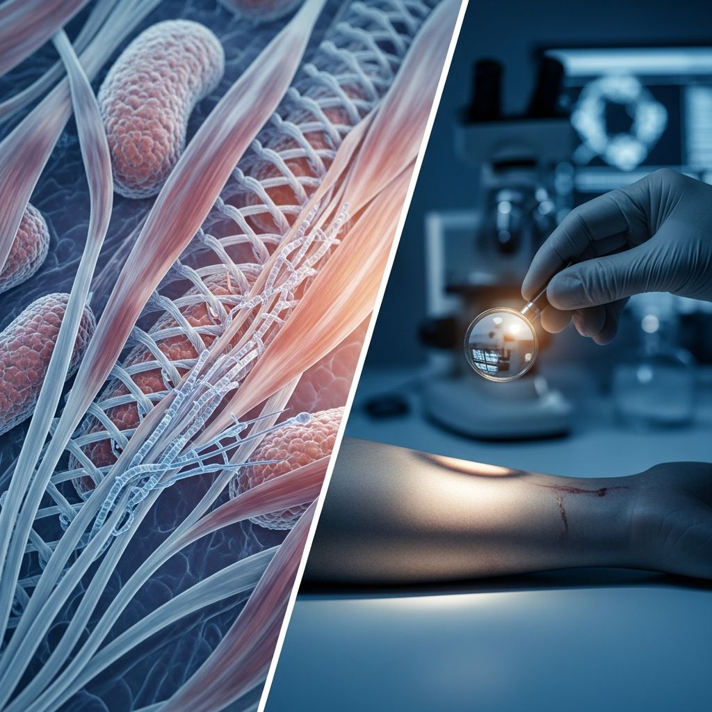
The Post-Mortem Transformation: Understanding Muscle Stiffening After Death
When a person dies, the human body undergoes a series of profound physical transformations. Among the most visible and scientifically significant is the phenomenon known as rigor mortis—the progressive stiffening of the body’s musculature that occurs in the hours following death. This phenomenon represents far more than a simple mechanical change; it reflects fundamental biochemical processes at the cellular level that forensic professionals use to help determine the approximate time of death and understand the circumstances surrounding a person’s passing.
Rigor mortis stands as one of four recognizable stages of post-mortem change, typically appearing several hours after the cessation of vital functions. Understanding the mechanisms behind this process requires knowledge of muscle physiology, cellular energy production, and the chemical interactions that normally allow muscles to contract and relax during life.
The Role of ATP: Cellular Energy and Muscle Function
At the heart of understanding rigor mortis lies a fundamental molecule: adenosine triphosphate, commonly abbreviated as ATP. During life, ATP serves as the primary energy currency of cells, providing the biochemical fuel necessary for virtually all cellular processes. In muscle tissue specifically, ATP plays a critical dual role—it enables muscle contraction and, equally importantly, it facilitates muscle relaxation.
The mechanism of muscle contraction involves two essential protein filaments: myosin and actin. These filaments interact through a complex dance of molecular binding and release. When a muscle receives a signal to contract, calcium ions flood into the muscle cell, triggering myosin and actin to bind together. This binding generates the mechanical force that produces muscle contraction. However, this bound state is not permanent. For the muscle to relax and return to its resting length, ATP must bind to the myosin filament, causing a conformational change that breaks the myosin-actin bond. Without this ATP-dependent release mechanism, the muscle remains locked in the contracted state.
Additionally, ATP powers calcium pump proteins embedded in the muscle cell membrane. These pumps actively transport calcium ions back out of the cell, restoring the electrochemical gradient necessary for normal muscle function and preventing unwanted re-stimulation of contraction. During life, these processes occur continuously and automatically, regulated by the nervous system and supported by the body’s aerobic respiration.
Aerobic Respiration Cessation and the Energy Crisis
The immediate consequence of death—whether from cardiac arrest, respiratory failure, or other causes—is the cessation of oxygen delivery to cells throughout the body. Without oxygen, cells cannot perform aerobic respiration, the metabolic pathway that efficiently generates ATP. In a living person, aerobic respiration in mitochondria produces approximately 30 to 32 ATP molecules from a single glucose molecule. This abundant energy production maintains all the electrochemical gradients and biochemical processes that keep muscles pliable and responsive.
Following death, this primary energy source vanishes. Cells retain only small residual stores of ATP, and the body’s muscles initially enter a state called primary flaccidity, in which all muscles relax completely. This occurs because the nervous system no longer sends electrical signals to muscles, and existing ATP reserves still permit normal myosin-actin dissociation and calcium reuptake. However, this relaxed state is temporary.
In the absence of oxygen, cells attempt to maintain minimal ATP production through anaerobic glycolysis—the metabolic breakdown of glucose without oxygen. This process, however, is dramatically less efficient than aerobic respiration, yielding only 2 ATP molecules per glucose molecule. Moreover, anaerobic metabolism produces lactate and pyruvate as byproducts, which accumulate in muscle tissue, creating an increasingly acidic intracellular environment.
Calcium Ion Dysregulation and Muscle Lock
As anaerobic ATP production gradually depletes the remaining cellular ATP stores, the calcium pumps begin to fail. These pumps require ATP energy to function, and without sufficient ATP, calcium ions cease being actively extruded from muscle cells. Simultaneously, calcium leaks from intracellular storage compartments into the sarcoplasm—the fluid within the muscle cell where myosin and actin filaments reside.
This influx of calcium reactivates the myosin-actin binding mechanism. The calcium binds to troponin, a regulatory protein on the actin filament, causing a conformational change that exposes the binding sites for myosin heads. Myosin and actin bind together, initiating muscle contraction. Under normal circumstances, this contraction would be brief and controlled, terminated by ATP binding to myosin. However, in the post-mortem state, ATP is virtually absent. The myosin heads remain bound to actin, unable to undergo the conformational change necessary for release. The muscle remains in a persistently contracted state.
The accumulation of lactic acid further reinforces this contracted state by lowering intracellular pH, which additionally impairs enzymatic processes that might otherwise help break these myosin-actin cross-bridges. The result is the characteristic rigidity of rigor mortis—muscles locked in contraction, unable to relax, gradually becoming visibly stiff to the naked eye.
Onset Timeline and Variable Factors
A common misconception about rigor mortis is that it appears immediately after death as a sudden, dramatic transformation. In reality, while the biochemical process begins instantly at the cellular level, visible stiffening typically requires several hours to become apparent. The timeline varies considerably based on numerous interconnected factors:
- Ambient temperature: Warm environments accelerate rigor mortis by increasing metabolic rates and hastening ATP depletion, while cold temperatures slow the process substantially.
- Physical condition: Individuals with greater muscle mass typically experience more pronounced rigor mortis, while persons with minimal muscle development may exhibit minimal or absent stiffening.
- Pre-mortem activity: Strenuous physical exertion immediately before death rapidly depletes ATP reserves, causing rigor mortis to develop more quickly. Similarly, convulsions, muscle spasms, or seizure activity immediately preceding death accelerates the process.
- Cause of death: Certain death mechanisms more rapidly deplete cellular ATP than others. Electrocution, for instance, causes rapid ATP exhaustion through severe muscle contractions.
- Systemic factors: Fever, infection, cerebral hemorrhage, severe illness, and certain poisonings (such as strychnine) all accelerate the development of rigor mortis.
- Substance use: Stimulant drugs including cocaine and amphetamine, which cause intense muscle activity, can accelerate rigor mortis onset.
- Age: Infants and newborns exhibit rigor mortis with exceptional rapidity, sometimes within minutes, while elderly individuals may show delayed onset.
In typical circumstances with moderate ambient temperatures and uncomplicated causes of death, visible rigor mortis usually appears between 2 and 6 hours post-mortem, though it may not be fully established for 12 hours or longer.
Sequential Progression Through Muscle Groups
Rigor mortis does not affect all muscles simultaneously. Instead, it follows a predictable anatomical sequence first systematically described in 1811 by French forensic physician Auguste Ambroise Tardieu and later codified as Nysten’s rule. This pattern begins with the involuntary muscles and progresses downward through the body in the following order:
- Heart muscles (cardiac muscle)
- Upper eyelids
- Neck muscles
- Jaw muscles
- Facial muscles
- Upper extremity muscles (arms and shoulders)
- Trunk muscles (chest and abdomen)
- Lower extremity muscles (legs and feet)
This cephalocaudal (head-to-toe) progression occurs because different tissues have varying ATP concentrations and metabolic rates. Tissues with higher initial ATP stores or lower metabolic demands resist rigor mortis longer. The heart, paradoxically, experiences rigor first despite being vital, likely due to its extremely high and continuous metabolic demands during life, leaving minimal ATP reserves upon death.
Understanding this progression assists forensic pathologists in estimating post-mortem interval, though numerous variables can modify this expected sequence.
Rigor Mortis Resolution and Advanced Decomposition
Rigor mortis is not permanent. As decomposition progresses, bacterial enzymatic action gradually breaks down the myosin-actin cross-bridges. Additionally, muscle tissue itself begins to decompose, breaking down the structural integrity of the protein filaments involved in rigor. Bodies in advanced states of putrefaction lose the characteristic rigidity of rigor mortis. Forensic pathologists can distinguish between rigor-induced stiffness and stiffness from advanced decomposition by examining tissue degradation patterns and other post-mortem changes.
Forensic Applications and Investigative Significance
Rigor mortis serves as a valuable tool in forensic pathology for several investigative purposes. The degree and distribution of rigor mortis can help narrow the post-mortem interval—the time elapsed since death. By considering rigor mortis progression alongside other post-mortem indicators such as livor mortis (blood settling and discoloration) and algor mortis (body cooling), forensic experts develop more precise death time estimates.
Additionally, unusual positioning of rigor mortis can provide investigative leads. When a body is discovered in an anatomically improbable position—such as limbs held against gravity or joints bent beyond normal range—and rigor mortis has preserved this positioning, it suggests the body may have been moved or posed after death, which could indicate foul play or concealment of the corpse.
The pattern and presence of rigor mortis also helps pathologists understand the circumstances immediately surrounding death. The absence of rigor mortis in a fresh body might suggest very recent death, while its presence and degree of advancement provides temporal context.
Frequently Asked Questions
Q: Does rigor mortis occur in all bodies?
A: Rigor mortis occurs in virtually all bodies, though its visibility and intensity vary. Individuals with very low muscle mass may show minimal stiffening, and certain pathological conditions may affect its development. Additionally, bodies recovered from cold environments may show delayed or reduced rigor mortis.
Q: Can rigor mortis be reversed?
A: No, rigor mortis cannot be reversed during its active phase. The myosin-actin cross-bridges persist until enzymatic degradation breaks them down, which begins with decomposition. Attempting to forcefully straighten a limb in rigor mortis can cause the muscles to tear rather than release.
Q: How does water temperature affect rigor mortis in drowning victims?
A: Cold water significantly slows rigor mortis development and can preserve its appearance for extended periods. Bodies in very cold water may show minimal decomposition and persistent rigor mortis weeks or months after death, whereas warm water accelerates both rigor mortis onset and subsequent decomposition.
Q: Why do some bodies show unusual positions in rigor mortis?
A: If a body is moved or positioned after death but before rigor mortis fully develops, the muscles will stiffen in that new position. Alternatively, intense muscular activity immediately before death can cause contractions that lock the body in unusual positions, such as the “pugilistic stance” seen in bodies exposed to extreme heat.
Q: Can forensic experts determine cause of death from rigor mortis patterns?
A: While rigor mortis timing and distribution can suggest certain types of deaths (such as poisonings or convulsions), it cannot definitively establish cause of death. Rigor mortis patterns must be interpreted alongside other forensic evidence, autopsy findings, and investigative information.
References
- Rigor mortis | Description, Cause, Mechanism, & Stages — Britannica. Accessed 2026. https://www.britannica.com/science/rigor-mortis
- What is rigor mortis, and why does it happen? — Live Science. https://www.livescience.com/health/what-is-rigor-mortis-and-why-does-it-happen
- Rigor mortis in an unusual position: Forensic considerations — National Center for Biotechnology Information (NCBI). https://pmc.ncbi.nlm.nih.gov/articles/PMC3657962/
- Everyday Q&A: What causes rigor mortis? — Yale Scientific Magazine. 2010. https://www.yalescientific.org/2010/02/everyday-qa-what-causes-rigor-mortis/
- Rigor mortis — Britannica. Accessed 2026. https://en.wikipedia.org/wiki/Rigor_mortis
- Rigor Mortis: Overview, Causes, 6 Stages, Duration, Death — Medicine Net. https://www.medicinenet.com/what_are_the_stages_of_rigor_mortis/article.htm

Medha Deb is an editor with a master's degree in Applied Linguistics from the University of Hyderabad. She believes that her qualification has helped her develop a deep understanding of language and its application in various contexts. Medha specializes in the areas of beauty, health, sports, and wellness and is committed to ensuring that the content on the website is…